
Intra-thoracic Idiopathic spinal cord herniation diagnosed with MRI and CT Myelography studies
Ventral cord herniation, also known by a variety of other terms such as spontaneous thoracic cord herniation or idiopathic spinal cord herniation, is a rare cause of focal myelopathy due to herniation of the thoracic cord through a dural defect.
Post surgical cord herniation can occur at any level, usually dorsally in the cervical region following laminectomy. The remainder of this article deals with spontaneous ventral thoracic herniation.
Epidemiology
In the majority of cases no possible antecedent cause is identified and the dural defect is thought to be congenital or idiopathic in nature. In a number of patients a history of previous trauma or surgery in the thoracic region may be present.
Clinical presentation
Clinical presentation is variable, generally with features of a myelopathy. However, progressive Brown-Sequard syndrome is classical, due to herniation of only one side of the cord.
Pathology
The underlying cause of ventral cord herniation is thought to be a dural defect allowing the subarachnoid space to communicate with the extradural space, which can be congenital or acquired The thoracic cord, naturally closely applied to the ventral dura due to the normal thoracic kyphosis then ‘plugs’ the hole and gradually herniates through the defect. It is this normal anatomical relationship between the ventral theca and anterior thoracic cord which accounts for this entity only being encountered in the anterior aspect of the mid to upper thoracic spine. The distortion of the cord parenchyma, formation of adhesions and possible vascular compromise in turn leads to myelopathy and neurological dysfunction.
Radiographic features
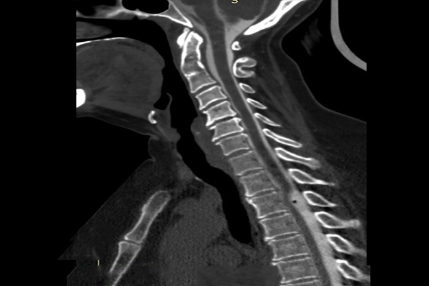
The key finding is focal distortion of the cord with a focal defect in the dura, and localised cord signal change. Although both CT and conventional myelography are able to visualise the distortion, MRI is the modality of choice to assess cord signal change and to evaluate for the differential diagnoses of spinal arachnoid cyst and dorsal thoracic arachnoid web.
MRI
Ventral cord herniation is only encountered between T2 and T8 where the normal thoracic kyphosis leads to the thoracic cord being in close proximity to the ventral theca.
The key feature is focal distortion and rotation of the cord with no CSF seen between it and the ventral theca. In most instances the cord is seen to bulge beyond the confines of the theca and is associated with T2 signal abnormality at that level.
Small extradural CSF intensity collection may also be seen, thought to represent the bulging CSF-filled arachnoid layer.
Treatment and prognosis
Surgery with division of adhesions and closure of the dural defect, which may require a dural graft/duroplasty, is curative. In most cases symptoms improve, however depending on the degree of pre-operative myelopathy complete recovery may not occur. Prominent T2 signal change within the cord may be a poor prognostic factor for full recovery .